A few months ago, I published a post about why inducing meiosis is important. Here, I will provide a short update about my preliminary efforts on this project.
The overall approach I am taking to induce meiosis is:
Performing a literature search and computational analysis to identify proteins involved in meiosis.
Constructing a barcoded transposon library for inducible expression of these proteins.
Integrating the library into iPSC lines with reporters for meiosis-specific protein
Inducing expression and sorting reporter-positive cells. Haploid cells could also be sorted by staining for DNA content, although this has a smaller dynamic range than fluorescent reporters.
Sequencing barcodes to identify the top factors.
Validating the top factors by expressing them individually or in small combinations.
Prediction of meiosis-inducing factors
To begin, my research assistant and I performed a literature search for meiosis-specific genes. We particularly focused on genes known to be involved in the initiation of meiosis. This search identified a total of 260 genes. Out of these, 14 RNA-binding proteins and transcription factors (TFs) were promising as regulatory factors.
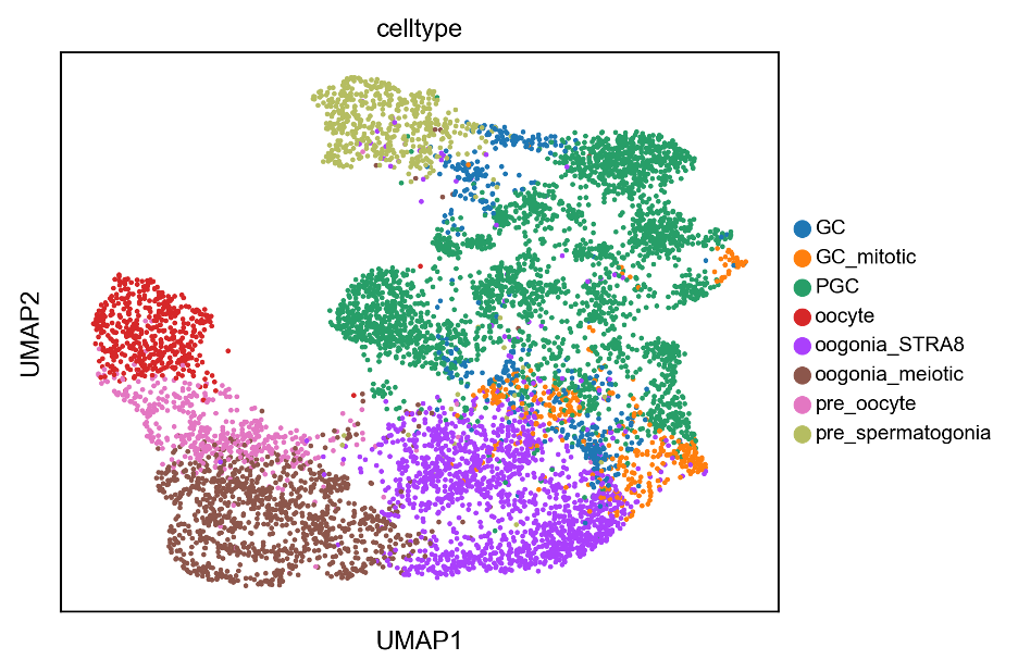
In parallel, I performed a pySCENIC analysis using a recently published scRNAseq dataset of human fetal gonads. The dataset contained germ cells in all stages of development: mitotic, premeiotic, meiotic, and postmeiotic.

pySCENIC constructs a gene regulatory network, then filters the network based on known TF binding motifs, and finally computes a score for each TF regulon in each cell. From this analysis, I identified 10 TFs important for premeiotic cells and 11 TFs for meiotic cells.
In addition to the pySCENIC TF predictions, I also chose a few additional TFs based on the gene regulatory network predicting them to regulate expression of the genes we identified in the literature search. I included these additional genes because sometimes pySCENIC has false negatives when TF binding motifs are not well-annotated.
In total, we identified 51 genes to screen for inducing meiosis. If you are a potential collaborator and want to see this list, please email me: metacelsus at protonmail dot com
Transposon Plasmid Construction
We are now constructing all the expression plasmids for these genes. We have completed 21 so far, and we will probably have all of them ready before the end of October. The final product will be a barcoded library of transposon plasmids containing our cDNAs under control of a doxycycline-inducible promoter.
Reporter iPSC construction
All this work generating and screening the library will be useless if we can’t tell whether or not we’re inducing meiosis. The naïve way to do this would be to see if cells have half their normal DNA content. However, this has several problems:
The dynamic range is at most 2, which is not very good.
It’s all-or-nothing: there’s no way to see that cells successfully enter meiosis unless they also complete meiosis.
It has a high false positive rate, because cellular debris can look like cells with a smaller amount of DNA.
There is also the potential for false negatives. For example, oocytes don’t complete meiosis until after fertilization, so they never have a haploid amount of DNA.
So, we won’t rely on DNA content as our main screening readout. Instead, we will use fluorescent reporters for two meiosis-related proteins, REC8 and SYCP3.
REC8 is a meiosis-specific cohesin which is expressed in pre-meiotic and early meiotic cells.1 SYCP3 is the main component of the synaptonemal complex lateral element, which is absolutely critical for homologous chromosome pairing during meiosis. SYCP3 is highly specific for meiotic cells. In terms of timing, REC8 expression shows that cells are preparing for meiosis, and SYCP3 expression shows that cells are actually performing meiosis.
I already have a female iPSC line with a REC8-T2A-mGreenLantern fluorescent reporter knock-in allele that I made as part of my oogenesis project. For this meiosis project, we will additionally engineer a male iPSC line with this allele, since we want to perform screening in both male and female cells.
The SYCP3 reporter will require a different design. Because SYCP3 must tightly pack with itself to form filaments, it is very sensitive to structural alterations, especially at the C-terminus. I don’t think I’ll be able to get away with the same design as my REC8 reporter (which has a few extra amino acids dangling off the C-terminus from the T2A sequence). Structural alterations to SYCP3 can cause a dominant-negative effect by interfering with filament assembly even when wild-type SYCP3 is present. Therefore, I think it’s better to completely replace one allele of SYCP3 with a cDNA for a fluorescent protein. Mice having only one SYCP3 copy are fertile, so I think this won’t interfere too much with meiosis. It's also more convenient because we don’t have to spend the extra effort to get a homozygous clone.
As an alternative, I am also considering a fluorescent sensor for SYCP3 RNA using RADAR, a new way to sense RNAs using adenosine deaminases.2
Finally, we will also perform scRNAseq on cell populations expressing our library to get a broader view of the gene expression changes our factors are inducing.
Noticing the skulls
During the literature review, I noticed that I was not the first to have the general idea for this project. A Scientific Reports paper in 2016 screened the expression of 12 germ cell-related genes in human fibroblasts and mesenchymal cells grown in spermatogonial stem cell culture medium. These researchers reported that a subset of six factors caused the cells to “display meiotic germ cell-like features”. However, this paper has a few issues:
The main “germ cell-like feature” they report was that their cells formed clumps in culture. Germ cells do this, but it’s not very specific for germ cells.
Germ cell marker gene expression was much lower than in vivo (and the authors used misleading Y-axis scales to try to hide this). Still, they do get some upregulation relative to the starting cells.
They report SYCP3 expression as a marker of meiosis . . . but SYCP3 was one of the six genes that they added, so they’re just seeing that. (The same goes for VASA, also known as DDX4).
Their data showing that cells are haploid isn’t particularly convincing (it’s a big claim, and I would expect to see a full karyotype to support it, which they don’t have). They could be getting something sort of like meiosis, but producing aneuploid cells.
In my experience, Scientific Reports papers vary widely in quality, and this is one of the weaker ones. Still, it’s worth noting what we will do to make our project better:
We will have a more complete library of meiosis-related factors (some of theirs were related to pluripotency rather than meiosis). Plus, this library will be barcoded for pooled screening.
We will screen our library in more biologically relevant cell types (iPSCs and PGCLCs, rather than fibroblasts and mesenchymal cells).
We will have a better readout for activation of meiosis (fluorescent reporters and scRNAseq, rather than clump formation and total DNA content).
Hopefully this approach will be more successful.
Project timeline
We anticipate finishing library construction by the end of this month. Engineering the reporter iPSCs will probably take until the end of December (with much of this time spent on validation and quality control). We are planning to perform the initial screens in early 2023. For the next few months, I will also be working to get my ovarian organoid project through peer review, but after that’s done, I will make meiosis my main focus.
The RNA stops being expressed after meiosis is completed, but the protein persists in oocytes (for 40+ years in the case of humans!) It does begin to wear out after a while, though, which contributes to oocyte aneuploidy in older women.
Yet another option is a promoter reporter rather than a knock-in reporter . . . but in my experience, promoter reporters aren’t great in terms of false positives and false negatives.


It might be cleaner to use an IRES sequence right after the stop codon of SYCP3 so your fluorescent reporter expresses off the same transcript without being conjugated directly to SYCP3. Would be an issue if you were imaging for SYCP3 localization, but you just want flow sorting, right?
It's a pretty well-known technology and I don't know the literature very well for it, but a bit of searching was able to find me this: https://www.ncbi.nlm.nih.gov/pmc/articles/PMC2275123/